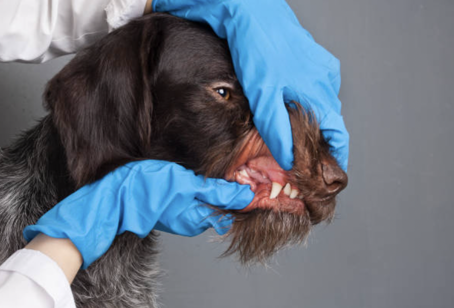
When it comes to veterinary dental procedures, extracting the maxillary fourth premolar in cats and dogs can be particularly challenging. Known as the maxillary carnassial tooth, this tooth (Triadan number 108, 208) is essential for slicing through flesh and is often deeply rooted, making it prone to fractures. These fractures can range from simple enamel cracks to more severe cases where the pulp is exposed, necessitating either intricate endodontic therapy or extraction.
The maxillary carnassial tooth, when fractured, typically suffers from what’s known as a slab fracture, affecting its largest cusp and often resulting in significant loss of enamel and dentin. If the fracture doesn’t expose the pulp, it’s considered an uncomplicated crown fracture, which might not require immediate treatment. However, more severe fractures that expose the pulp or extend beneath the gum line complicate the situation, often requiring surgical intervention.
For those in the veterinary field, or even pet owners interested in understanding more about this common issue, here’s a detailed guide on how to handle the extraction of a firmly rooted maxillary fourth premolar, minimizing complications and ensuring the best outcome for our furry friends.
Step-by-Step Guide to Extracting the Maxillary Fourth Premolar
1. Preparing for Extraction:
– Begin with mesial and distal releasing incisions connected by a sulcular incision using a sharp scalpel blade. This helps in freeing the attached gingiva.
– It’s crucial to be aware of nearby anatomic structures such as the neurovascular bundle near the infraorbital foramen to avoid accidental damage.
2. Lifting the Gum:
– Use a periosteal elevator to gently lift the mucoperiosteal flap. Keeping your instruments sharp will aid in efficiently separating the gum from the bone without unnecessary tearing.
3. Accessing the Root:
– Employ a #2 surgical round bur to create windows in the bone adjacent to the tooth roots. This step is vital for reducing the amount of bone removal needed, preserving the structural integrity of the jaw.
4. Sectioning the Tooth:
– With a crosscut fissure bur, carefully section the tooth into three parts. This technique helps in isolating the segments and makes them easier to remove individually.
5. Separating the Tooth Segments:
– Test the separation by using a dental elevator in the created spaces. Ensuring each segment is free before attempting extraction is key to a smooth procedure.
6. Removing the Tooth:
– Gradually work the tooth segments loose using a combination of elevators and luxators, tools designed to cut through the periodontal ligament and leverage against the alveolar bone.
7. Final Steps:
– Once the segments are loose, use extraction forceps to gently remove them from the socket. Be patient and apply gentle force to avoid breaking the tooth further.
8. Post-Extraction Care:
– Debride and lavage the socket to clean out any debris, and smooth any sharp bone edges to prevent discomfort as the area heals.
– Obtain a postoperative radiograph to confirm that all segments of the tooth have been successfully removed.
9. Closing the Site
– Ensure the flap is not under tension when sutured. A tension-free closure promotes better healing and reduces the risk of complications.
This comprehensive approach not only ensures the safe removal of a problematic tooth but also preserves as much of the surrounding tissue as possible, leading to better outcomes for the pet. Whether you’re a seasoned vet or a student, understanding and mastering these steps can significantly enhance your skills in veterinary dentistry.


